


 Book Now
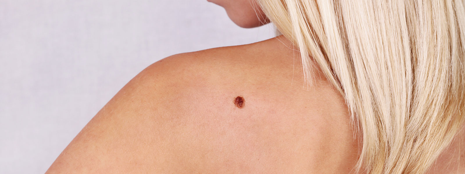
MOHS Micrographic Surgery allows the selective removal of areas of skin affected by skin cancer while protecting as much normal tissue as possible.
This method of surgery is named after the physician, Frederic E. Mohs, MD, who developed this surgical technique. Even though this technique is time consuming, the accuracy and chance for the complete removal of skin cancer is very good. As a result, MOHS micrographic surgery is very useful for large or aggressive tumors, those with indistinct borders, near vital functional or cosmetic structures and for tumors where other therapies have failed.
Mohs surgeons are dermatologists who have performed training in Mohs micrographic surgery.
Indications
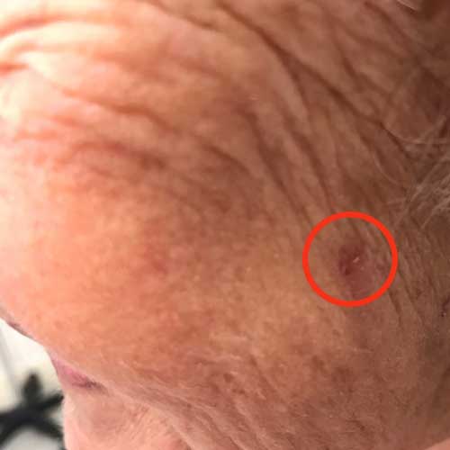
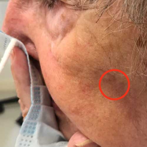
The majority of tumors treated with Mohs Micrographic surgery are complex basal and squamous cell carcinomas.
Skin cancers are complex when:
- • Cancer is in an area where preservation of healthy tissue is critical to maximize function and cosmetic result (eyelids, nose, ears, lips, hands)
- • Cancer is in an area of higher tumor recurrence (ears, lips, nose, eyelids, temples)
- • Cancer was incompletely treated, or was previously treated and is recurrent
- • Cancer is large
- • The edges of the cancer cannot be clearly defined
- • Scar tissue exists in the area of the cancer
- • Cancer grows in an area of prior radiation therapy
- • The patient is immunosuppressed (organ transplant, HIV infection, chronic lymphocytic leukemia)
- • The patient is prone to getting multiple skin cancers (including genetic syndromes such as basal cell nevus syndrome and xeroderma pigmentosa)
The MOHs Surgery Procedure
The Mohs surgical process involves a repeated series of surgical excisions followed by microscopic examination of the tissue to assess if any tumor cells remain. Some tumors that appear small on clinical exam may have extensive invasion underneath normal appearing skin, resulting in a larger surgical defect than would be expected. It is therefore impossible to predict a final size until all surgery is complete. As Mohs surgery is used to treat complex skin cancers, approximately half of all treated tumors require 2 or more stages for complete excision.
MOHs Surgery steps in detail:
Step 1: Anesthesia
The tumor site is locally infused with anesthesia to completely numb the tissue. General anesthesia is not required for Mohs micrographic surgery.
Step 2: Stage I – Removal of visible tumor
Once the skin has been completely numbed, a thin saucer-shaped “layer” of tissue is surgically removed by the Mohs surgeon. An electric needle may be used to stop the bleeding.
Step 3: Mapping the tumor
Once a “layer” of tissue has been removed, a “map” or drawing of the tissue and its orientation to local landmarks (e.g. nose, cheek, etc) is made to serve as a guide to the precise location of the tumor. The tissue is labeled and color-coded to correlate with its position on the map. The tissue sections are processed and then examined by the surgeon to thoroughly evaluate for evidence of remaining cancer cells. It takes approximately 60 minutes to process, stain and examine a tissue section. During this processing period, your wound will be bandaged and you may leave the operative suite.
Step 4: Additional stages – Ensuring all cancer cells are removed
If any section of the tissue demonstrates cancer cells at the margin, the surgeon returns to that specific area of the tumor, as indicated by the map, and removes another thin layer of tissue only from the precise area where cancer cells were detected. The newly excised tissue is again mapped, color-coded, processed and examined for additional cancer cells. If microscopic analysis still shows evidence of disease, the process continues layer-by layer until the cancer is completely removed.
This selective removal of tumor allows for preservation of much of the surrounding normal tissue. Because this systematic microscopic search reveals the roots of the skin cancer, Mohs surgery offers the highest chance for complete removal of the cancer while sparing the normal tissue. Cure rates typically exceed 99% for new cancers, and 95% for recurrent cancers.
Step 5: Reconstruction
Reconstruction is individualized to preserve normal function and maximize aesthetic outcome. The best method of repairing the wound following surgery is determined only after the cancer is completely removed, as the final defect cannot be predicted prior to surgery. Stitches may be used to close the wound side-to-side, or a skin graft or a flap may be designed. Sometimes, a wound may be allowed to heal naturally.
Although no surgeon or technique can guarantee a 100% chance of cure, this surgical technique offers a 97-99% chance for the complete removal of skin cancers.